• IP addresses are NOT logged in this forum so there's no point asking. Please note that this forum is full of homophobes, racists, lunatics, schizophrenics & absolute nut jobs with a smattering of geniuses, Chinese chauvinists, Moderate Muslims and last but not least a couple of "know-it-alls" constantly sprouting their dubious wisdom. If you believe that content generated by unsavory characters might cause you offense PLEASE LEAVE NOW! Sammyboy Admin and Staff are not responsible for your hurt feelings should you choose to read any of the content here.

    The OTHER forum is HERE so please stop asking.

Collateral Circulation is the reason you need regular exercise. It may save you one day when your main artery is blocked

SBFNews

Alfrescian
Loyal
my.clevelandclinic.org

What is collateral circulation?​

Collateral circulation is alternate or “backup” blood vessels in your body that can take over when another artery or vein becomes blocked or damaged. Your collateral circulation provides alternative routes for blood flow.

Cleveland Clinic is a non-profit academic medical center. Advertising on our site helps support our mission. We do not endorse non-Cleveland Clinic products or services. Policy

How does collateral circulation work?​

Collateral blood vessels connect to your main blood vessels and remain small and unused until you need them. Other collateral vessels develop throughout life in a process called angiogenesis (the formation of new blood vessels).

Angiogenesis may happen as a main blood vessel slowly becomes blocked. Your body senses the reduced blood flow and calls other blood vessels into action. Regular aerobic exercise, such as running, may also encourage new collateral blood vessels to form.

How common is collateral circulation?​

Usually, people are born with some collateral blood vessels, but the number and size are different for each person. Collateral blood vessels can be:
  • Arteries, larger blood vessels that carry blood away from your heart.
  • Capillaries, tiny blood vessels that help transport oxygen and nutrients.
  • Veins, carry blood back to your heart.

What health conditions are related to collateral circulation?​

Your collateral circulation may take over if you have health conditions that restrict blood flow, such as:
Collateral blood vessels cannot always prevent ischemia (lack of blood supply) from these health conditions. Collateral vessels must expand quickly enough to take over blood flow before the blood supply is completely blocked. These health conditions always require treatment, even in people who have good collateral blood vessels.

How long does collateral circulation last?​

When your collateral circulation takes over, smaller blood vessels must get larger to accommodate the increased blood flow. The enlarged collateral vessels may continue to accommodate your normal blood flow for months or years.
But as you get older, your collateral blood vessels gradually shrink. This process, called rarefaction, is more likely to happen in people who have cardiovascular disease. When your collateral blood vessels shrink, they are less likely to work, which can lead to symptoms when blood flow becomes blocked in your main blood vessels.

Where does collateral circulation occur?​

Various parts of your body can have collateral circulation, including your:
  • Abdomen.
  • Arms.
  • Brain.
  • Feet and legs.
  • Heart.
  • Lungs.
A note from Cleveland Clinic
Collateral circulation is your body’s way of working around a blockage in blood flow. It’s not a foolproof way to prevent damage from conditions like stroke and PAD, or to prevent a heart attack or stroke. However, it can be helpful and possibly lifesaving in some cases.

Researchers are continuing to study collateral circulation and the possibility of creating and using artificial collateral veins. This could expand the body’s natural ability to overcome a variety of conditions that obstruct blood flow.
 

SBFNews

Alfrescian
Loyal

Coronary Collateral Growth Induced by Physical Exercise: Results of the Impact of Intensive Exercise Training on Coronary Collateral Circulation in Patients With Stable Coronary Artery Disease (EXCITE) Trial​


Background: A well-developed coronary collateral circulation provides a potential source of blood supply in coronary artery disease. However, the prognostic importance and functional relevance of coronary collaterals is controversial with the association between exercise training and collateral growth still unclear.

Methods and results: This prospective, open-label study randomly assigned 60 patients with significant coronary artery disease (fractional flow reserve ≤0.75) to high-intensity exercise (group A, 20 patients) or moderate-intensity exercise (group B, 20 patients) for 4 weeks or to a control group (group C, 20 patients). The primary end point was the change of the coronary collateral flow index (CFI) after 4 weeks. Analysis was based on the intention to treat. After 4 weeks, baseline CFI increased significantly by 39.4% in group A (from 0.142±0.07 at beginning to 0.198±0.09 at 4 weeks) in comparison with 41.3% in group B (from 0.143±0.06 to 0.202±0.09), whereas CFI in the control group remained unchanged (0.7%, from 0.149±0.09 to 0.150±0.08). High-intensity exercise did not lead to a greater CFI than moderate-intensity training. After 4 weeks, exercise capacity, Vo2 peak and ischemic threshold increased significantly in group A and group B in comparison with group C with no difference between group A and group B.

Conclusions: A significant improvement in CFI was demonstrated in response to moderate- and high-intensity exercise performed for 10 hours per week.

https://pubmed.ncbi.nlm.nih.gov/26979085/
 

SBFNews

Alfrescian
Loyal
Natural Bypasses Can Save Lives
www.ahajournals.org

Blood to the heart muscle is provided by the left and right coronary arteries, which arise from the aorta and then branch into increasingly tinier vessels.

Each coronary artery supplies blood to its own area of muscle (Figure 1). In humans, very small, hairlike vessels (capillaries) are often the only interconnections between the coronary arteries and their service areas. Sometimes, however, larger vessels interconnect the supplied areas. These vessels are called “collateral vessels” or “natural bypasses.” Only these collaterals, not the capillary network, are capable of delivering an adequate amount of blood to the heart muscle. The extent of these natural bypasses varies from person to person and depends on individual conditions, such as hereditary factors, the degree of physical activity, and the existence of constrictions in the main coronary arteries, called “coronary artery disease.”

2ff1.jpg
Figure 1. Influence of collateral supply on infarct size. A, Occlusion of a coronary artery with subsequent myocardial infarction. Before the occlusion occurred, the area of infarction (depicted in gray) was not supplied sufficiently by collateral vessels; thus, the size of the infarct comprises a large area (that is, the entire area at risk). B, Occlusion of the same coronary artery at the same site, but in this case, the area at risk was well supplied by collateral vessels (arrows). Therefore, this patient will have no infarction at all.

Myocardial Infarction Is a Major Cause of Death​

In industrialized countries, coronary artery disease is a major killer. The disease is characterized by a general inflammatory process of the entire coronary artery system, with development of local deposits (called “plaques”) of lipids, cells of inflammation, connective tissue, and calcium. This can lead either to progressively slow narrowing of the affected artery (which is typically sensed as angina pectoris) or a sudden rupture of a plaque (Figure 1) caused by its instability. In the latter situation, the downstream blood flow is abruptly blocked (which is the classic “heart attack” or myocardial infarction; Figure 1).

Factors That Determine the Size of an Infarct​

As a consequence of such a blockage, the section of heart muscle normally supplied by the vessel lacks “fuel,” that is, oxygen and nutrients, and it finally dies. If the patient survives, the final stage is a loss of cardiac pumping strength and a scar that replaces the former muscle. The size of an infarction is not the same in every case: It is mainly influenced by the size of the blocked artery (that is, a larger artery supplies a larger area of muscle, the so-called area at risk), the duration of the blockage (that is, the damage is smaller when rapid reopening of the artery occurs), and importantly, the extent of blood flow through collaterals from neighboring vessels (that is, the more that collaterals can “step into the breach” to supply the starving muscle tissue in need, the smaller is the size of the infarction; Figure 2). In the ideal case, a very good collateral supply to the area at risk can make the infarct size shrink to zero when there is a sudden blockage of an artery. In the worst case, when no or a very low collateral flow to the starving area of infarction is available, the infarct involves the entire area at risk of the supplying vessel, which is blocked.

2ff2.jpg
Figure 2. Coronary angiography with an example of the muscle- and life-saving impact of collaterals. In this patient, the right coronary artery (RCA) is filled with contrast agent. By a natural bypass, that is, the collateral channel (arrows), the left coronary artery (LAD), which is blocked at its origin (ring), is filled with blood. Without the collateral vessel providing blood to the large territory of the left coronary artery, this area would have died off; that is, the patient would have suffered a potentially deadly infarction.

Established Treatments for Coronary Artery Disease​

Besides treatment with drugs, constricted vessels can be widened by inflating a balloon on a catheter (angioplasty) and, in most cases, also placing a wire tube (stent) to keep the vessel open. In an infarction, this is the superior therapy, because blood flow to the heart muscle is restored as quickly as possible, and damage can be limited. Coronary artery bypass grafting (CABG; pronounced “cabbage”) operation is needed if numerous severe constrictions near the outlets of the coronary arteries are present. Altogether, these technologies have led to enormous progress in the treatment of the disease in the last 30 years.

Treatment Limitations​

As in every catheter-based intervention or surgical operation, there are risks related to the procedures. Apart from that, approximately one fourth of all patients are not suitable for these revascularization procedures at all. These procedures do indeed repair the local problem of narrowings, but they do not alter the natural progression of the disease. Alternative treatment practices are therefore required to alter the course of coronary artery disease, alleviate angina pectoris, and thereby decrease the number of deaths due to infarction.

How to Turn the Capillary Network Into Collaterals

When there is increasing traffic volume on a highway, it may make sense to make the highway into a larger freeway to allow a higher traffic volume. In short, the same happens to the coronary arteries: When blood flow is increased, the inner layer of vessel cells (endothelial cells) sense this necessity and start the process of enlarging from capillaries into genuine collateral vessels. In response to endurance exercise training (such as running, bicycling, swimming, and hiking), blood flow is increased, which leads to a conversion from capillaries into collaterals. This is a very elegant treatment everybody can accomplish. It reduces the chances of the occurrence of angina pectoris, myocardial infarctions, and death. Beyond the interventional, surgical, and medical treatments against coronary artery disease, this collateral training is a natural and valuable therapy that many patients can apply by themselves, for themselves, if only they are aware of it.

Key Points​

  • As a pump, the heart muscle is critically dependent on the coronary arteries for sufficient blood supply.
  • Narrowing of the coronary arteries by deposits (plaques) can starve the heart muscle of oxygen and nutrients. This results in chest pain (angina pectoris).
  • A heart attack results when a diseased coronary artery is blocked completely. As a consequence, the portion of the heart muscle normally supplied by this artery dies (myocardial infarction).
  • After a myocardial infarction, a certain mass of muscle is lost forever. Depending on infarct size, quality of life and life expectancy can be shortened.
  • Collateral training results in a better blood supply of the heart and thus demonstrably fewer heart attacks, less severe loss of valuable heart muscle, better quality of life, and higher survival rates.
The information contained in this Circulation Cardiology Patient Page is not a substitute for medical advice, and the American Heart Association recommends consultation with your doctor or healthcare professional.
 

SBFNews

Alfrescian
Loyal
'Back-up system' reduces heart disease deaths | UCL News
www.ucl.ac.uk


Small bypass vessels which act as a 'back-up system' for the heart's main arteries play a significant role in reducing the mortality of patients with coronary artery disease, according to new research.
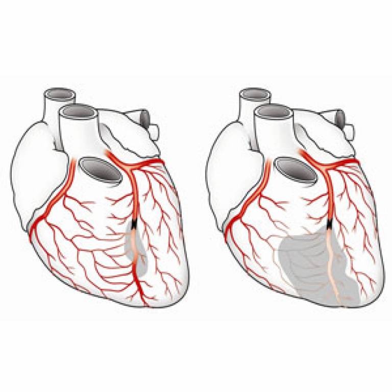
Illustration of coronary collaterals
Researchers from UCL, University of Bern, Yale University and other international collaborators examined the role of natural bypass vessels called coronary collaterals in patients with blocked arteries. The study, published online today in the European Heart Journal, shows that patients with lots of these vessels have a 36% reduced risk of mortality, highlighting their importance as a therapeutic target.

Coronary collaterals are tiny, specialised blood vessels that connect the larger vessels in the heart. They can be thought of as the heart's 'back-up system' as they are essentially invisible until activated, when they can enlarge their diameters in order to carry significant blood flow and bypass blockages. For many years, doctors believed that there were no connections between the main coronary arteries, and if one of these arteries got blocked patients would normally undergo bypass surgery or stenting. However, research is now building up concerning the importance of coronary collaterals.

This study pooled data from 12 studies enrolling 6,529 patients.

Researchers compared patient survival rates in participants with a high number of natural bypass vessels with those with minimal bypass vessels. Survival rates were higher among those who had a higher number of well developed vessels, compared to those with fewer or no such vessels. It is not yet clear why some people have better bypass networks than others, but scientists believe genes and lifestyle factors play an important role.

Lead author Dr Pascal Meier, a consultant at The Heart Hospital (part of University College Hospitals NHS Foundation Trust) and scientist at UCL Institute of Cardiovascular Science, commented: "When we see patients with a heart attack (clotted coronary artery), the damage caused to the heart varies greatly from person to person. One reason for this is because the 'back-up system' is better developed in some patients - but our study is the first to clearly show a difference in mortality. We found that whether patients underwent stenting to open their blocked artery or were treated with medications only, they had improved survival if their natural bypass vessels were better developed.
"A growing body of research demonstrates that these vessels are protective and reduce mortality in patients with blocked coronary arteries. We should find means to promote these natural bypass vessels in order to improve outcomes for patients with heart disease."

Dr Christian Seiler, Professor of Cardiology at the University Hospital Bern and senior investigator of this study, said: "We know that regular physical activity can improve the natural bypass network. Recently, some small studies have examined possible ways of promoting natural bypass vessels, such as a treatment known as external counterpulsation (which imitates physical activity) and injections with a growth factor called G-CSF. Our study adds weight to the increasing evidence that we should be focusing more of our efforts on identifying how we can better develop natural bypass vessels."

Dr Pascal Meier was appointed as a result of the Yale-UCL Collaborative and will continue to work closely with Yale scientists to develop new cardiovascular devices and treatments.

Image: The left figure shows a heart with well-developed natural bypass vessels (coronary collateral arteries), the right figure shows a heart with poorly developed natural bypass vessels, leading to a much larger area with lack of blood supply (grey) in case of a blockage.
Media contact: Ruth Howells


Links:
Dr Pascal Meier
Yale UCL Collaborative

UCL Institute of Cardiovascular Sciences
University of Bern
 

glockman

Old Fart
Asset
KNN, after reading the various articles and watching the videos, I think I can start to treat and advise patients. Expert liao.:biggrin:

In a nutshell, exercise and keep fit. What doesn't kill you will make you stronger!:thumbsup:
 

SBFNews

Alfrescian
Loyal
KNN, after reading the various articles and watching the videos, I think I can start to treat and advise patients. Expert liao.:biggrin:

In a nutshell, exercise and keep fit. What doesn't kill you will make you stronger!:thumbsup:
Yep, must aim for 85% to 95% of Maximum Heart Rate a couple of times a week so that natural bypass vessels are developed. Everyone's main arteries will sooner or later be clogged up as we age. Calcium deposits build up over timr. So it's important to develop natural bypass vessels as a backup system.
 
Top